Conduits for Siliceous Opal-Precursor in Copepod Jaws
Conduits for Opal-Precursor Silicate in Copepod Jaws
Chapter 12 of
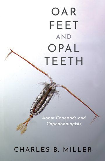
Oar Feet and Opal Teeth covers egg to adult development of copepods. It includes a section on how stone-like teeth, clasped like jewels in chitin bezels, are placed on the
outside of the mandibular exoskeleton. Long ago, coauthors and I wrote a paper1 to summarize our work toward understanding the process. Recently, while discarding old files, I found some transmission electron micrographs (TEMs) we did not publish. Perhaps that was because of some displayed gaps in tissue preservation.
However, the sections for these TEMs cut along the cores of the “duct” we believed carries a silica-bearing substance from a gland at the base of the mandible to the fibrous tooth molds on the developing jaw edge (Figure 12.19B in Oar Feet and Opal Teeth). Duct is in quotes, because there seems to be no actual lumen. None of the other micrographs with shorter lengths of opal-precursor duct showed a lumen.
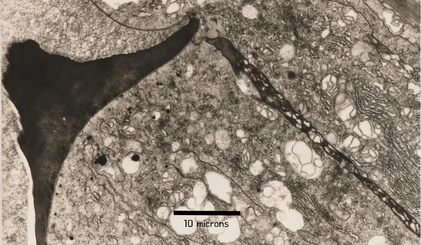
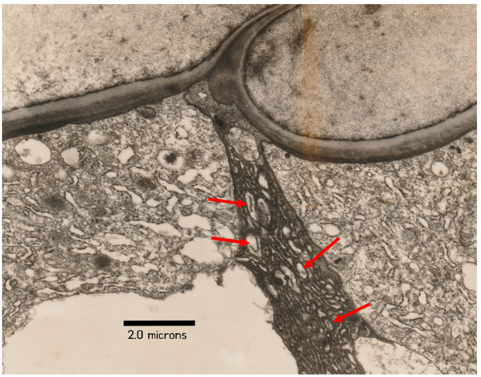
The rediscovered TEMs (Figures A & B) show the ducts’ distinctive structure of osmophilic (osmium staining) “lamellae.” I’ll call them “conduits.” They appear to have been somehow moving oval inclusions toward the tooth molds at the moment of preservation. Those blebs would be very difficult to characterize biochemically; they are smaller than many single bacteria. However silicious transporter proteins are known from diatoms2, rice and other plants.
Animals other than copepods have siliceous hard parts. Radulae of snails and chitons have hard, siliceous foundations. Those can have caps of harder hematite. Tissues of plants, including grasses and
Equisetum, have siliceous structures. Do the silica deposition systems in any other organisms incorporate opal-precursor conduits resembling those shown here from copepods?
If anyone would like to initiate a new study of tooth formation in copepods, they should know the barriers to catching ‘the process in process.’ Ask me at the email address given in “contacts.”
Regards, Charlie Miller
1. Miller, C.B., D.M. Nelson, C. Weiss & A.H. Soeldner (1990) Morphogenesis of opal teeth in calanoid copepods. Marine Biology 106: 91-101.
2. Knight, M.J. et al. (2016) Direct evidence of the molecular basis for biological silicon transport. Nature Communications |DOI:10.1038/ncomms11926 (11 pp.)
======================= Figures below=======================
Figure A. Neocalanus plumchrus, fifth copepodite.
TEM of a section near the developing tooth row prior to molting. It shows a nearly finished adult tooth at left, and at right the long opal-precursor conduit from the gland at the base of the mandibular gnathobase. The length of duct included in this TEM was about 45 microns. Not all of the cellular ultrastructure is well defined, likely due to bad tissue fixation. Fixative applied to live copepods does not readily penetrate deeply below the chitin exoskeleton.

Figure B. Neocalanus plumchrus, fifth copepodite
More enlarged view of an opal-precursor “duct” before it passes around curving sections of new exoskeleton toward a tooth “mold” on the jaw’s cutting edge. Red arrows indicate oval inclusions that possibly are an opal precursor.

Ways to Share

Oar Feet and Opal Teeth

